Genome sequence and gene
compaction of the eukaryote
parasite Encephalitozoon cuniculi
Michaël D. Katinka*, Simone Duprat*, Emmanuel Cornillot**, Guy Méténier**, Fabienne Thomarat**, Gérard Prensier**, Valérie Barbe*, Eric Peyretaillade**, Philippe Brottier*, Patrick Wincker*, Frédéric Delbac**, Hicham El Alaoui**, Pierre Payret**, Williarn Saurin*, Manolo Gouy§, Jean Weissenbach* & Christian P. Vivarès**
* Genoscope, UMR WRS 8030, CP 5706, 91057 Evry cedex, France
** Parasitologie Moléculaire et Cellulaire, Laboratoire de Biologie des Protistes, UMR CNRS 6023, Université Blaise Pascal 63177 Aubière cedex, France
§ Laboratoire
de Biométrie et Biologie Evolutive, UMR CNRS 5558, Université
Lyon I, 69622 Villeurbanne cedex, France
Microsporidia are obligate intracellular parasites infesting many animal
groups (1). Lacking mitochondria and peroxysomes, these unicellular eukaryotes
were first considered a deeply branching protist lineage(2) that diverged
before the endosymbiotic event that led to mitochondria. The discovery of
a gene for a mitochondrialtype chaperone (3-5) combined with molecular phylogenetic:
data (6-9) later implied that microsporidia are atypical fungi that lost mitochondria
during evolution. Here we report the DNA sequences of the 11 chromosomes of
the 2.9 megabase (Mb) genome of Encephalitozoon cuniculi (1,997 potential
proteincoding genes). Genome compaction is reflected by reduced intergenic
spacers and by the shortness of most putative proteins relative to their eukaryote
orthologues. The strong host dependence is illustrated by the lack of genes
for some biosynthetic pathways and for the tricarboxylic acid cycle. Phylogenetic
analysis lends substantial credit to the fungal affiliation of microsporidia.
Because the E. cuniculi genome contains genes related to some mitochondrial
functions (for example, Fe-S cluster assembly), we hypothesize that microsporidia
have retained a mitochondrion-derived organelle.
Encephalitozoon cuniculi infects various mammals, including humans, and can
cause digestive and nervous clinical syndromes in HIV-infected or cyclosporine-treated
people (1). Its reproduction proceeds as a sequence of two major stages: merogony,
involving the multiplication of large, wall-lacking cells (meronts); and sporogony,
leading to small, thick-walled spores. The sporal invasive apparatus is characterized
by a long polar tube that can be quickly extruded then used for transferring
the sporoplasm into the target cell. Consisting of 11 linear chromosomes ranging
from 217 to 315 kb, the E. cuniculi genome is remarkably reduced (-2.9Mb)
(10). The nucleotide sequence of the smallest chromosome has been recently
reported (11). The full sequencing of this minimal genome among eukaryotes
was expected to provide insight into the metabolism and general biology of
microsporidia and to help in the understanding of the evolutionary history
of amitochondriate eukaryotes currently considered 'curious fungi' (9) (see
Table 1).
The chromosome sequences were determined through a plasmid library (3 kb inserts)
and a miniBAC library (20-25 kb inserts) totalling 15 genome equivalents (46
Mb). All chromosomes possess a 'unique sequences' core region flanked by two
28-kb divergently oriented regions, each including one ribosomal DNA unit
(11,12). The subtelomeric repeats upstream from rDNA are mostly degenerated
minisatellites, whereas downstream repeats consist essentially of non-polymorphic
microsatellite arrays. The chromosome cores lack simple sequence repeats,
minisatellite arrays and known transposable elements. No imprint of retrogenes
or pseudoretrogenes of either polymerase (pol) II or pol III type was found.
General features of genome organization are indicated in Table 1.
Table 1 General features of the E. cuniculi genome
| Total sequenced length | 2,507,519 bp |
| G + C content | |
|
47.6% |
|
45.0% |
|
52.9% |
| No. of protein-coding sequences | 1,997 |
| Mean intergenic distance | 129bp |
| Gene density of chromosome cores | 1 CDS per 1,025 bp |
| No. and sizes of spliceosomal introns | 13 (23-52 bp) |
| No. of 16S-23S rRNA genes | 22* |
| No. of 5S rRNA genes | 3 (on chrV, VII, IX) |
| No. of tRNA genes | 44** |
| No. and sizes of tRNA introns | 2 (16, 42 bp) |
* Two per chromosome.
**On all chromosomes.
[fig. 1 graph not available]
The gradual increase in
G + C content at the core centre described in chromosome I (chrI)(11) exists
in all the chromosomes (maximum 51.0%). The 1,997 protein-coding DNA sequences
(CDSs) represent about 90% of the chromosome cores, as a result of generally
short intergenic regions (see Supplementary Information). Gene density is
slightly lower than that observed in the nucleomorph genome of the cryptomonad
Guillardia theta (13). Only about 44% of the CDSs are assigned to functional
categories and about 6% to conserved hypothetical proteins. In contrast to
the nudeomorph genome (13), no overlapping of CDSs with predicted functions
was revealed. Structural or functional clusters are rare and never composed
of more than two CDSs (for example, histones H3 and H4 on chrIX). Genome compaction
can also be related to gene shortening, as indicated by the length distribution
of all potential proteins (Fig. 2a). The mean and median lengths of all potential
E. cuniculi proteins are only 359 and 281 amino acids, respectively.
We compared the lengths of 350 proteins with Saccharomyces cerevisiae homologues
(Fig. 2b). More than 85% of these proteins are shorter than in yeast, with
a mean relative size difference of 14.6%. From the analysis of the protein
size distributions derived from sequenced genomes, it has been suggested that
the lengthening of proteins in eukaryotes (non-parasitic species) allows for
more complex regulation networks (14). Thus, protein shortening in E. cuniculi
may reflect reduced protein-protein interactions as a result of various gene
losses linked to the intracellular parasitic nature (Fig. 2a, b).
Perfect segmental duplications of 03-10-kb coding and noncoding sequences
represent about 3.7% of the core region in average (from 1% in chrII, III,
IV and VI to 7% in chrIX; see Supplementary Information). A segment carrying
four enzymecoding genes is perfectly duplicated in the extreme part of the
chrI core (11) but partially duplicated near the end of chrVIII (truncated
serine hydroxymethyltransferase gene). The genome core sequences exhibit a
very low base polymorphism, restricted to a few positions in eight chromosomes.
A duplicated gene homologous to CTP synthases (chrXI) revealed a rare polymorphic
tract of 189 base pairs (bp) (116,847-117,036). Hybridization experiments
indicate that each polymorphic sequence is present in about 50% of the DNA
molecules (data not shown), suggesting that they are alleles and supporting
the diploidy hypothesis (10, 12).
A large proportion of CDSs is assigned to the conservation and transmission
of genetic information as well as to protein modification and intracellular
transport processes, but with a significant degree of simplification, mainly
related to the lack of DNA-containing organelles. For example, subunits for
DNA pol I , pol II and pol III, are present while there is no candidate for
the mitochondrial DNA polymerase gamma. Apart from the telomerase catalytic
subunit, no RNA-dependent reverse transcrip tase was found. This and the lack
of any retrotransposition elements may explain the absence of pseudoretrogenes.
The potential transcription machinery includes RNA pol I, II and III, more
than 70 messenger RNA transcription-associated proteins and a virtually complete
set of genes for splicing, 5' and 3' processing. An (A + T)-rich consensus
transcription initiation sequence is revealed in the 120-bp region upstream
from the initiation codon of numerous genes, suggesting short 5' leaders.
Seventy different eukaryotic-type dbosomal proteins (40 from the large subunit,
30 from the small subunit) are predicted, which is slightly lower than in
the amitochondriate protist Giardia lamblia (74) (15). Compared with
the cytoplasmic ribosome of S. cerevisiae, the missing proteins are
LP1, L14, L29, L38, L40, L41, S27 and S27A. Small putative spliceosornal introns
with usual GT-AG boundaries were detected in 11 ribosomal protein genes (S8,
S17, S24, S26, LS, L19, L27A, L37, L37A and L39). They start either in the
initiator ATG or the next codon, as often observed in yeast (16) or nucleomorph
(13) genomes. Two more internal introns create frame shifts within a CDP-diacylglycerol
serine phosphatidyltransferase gene (chrXI). Two of the 44 transfer RNA genes
(tDNAile and tDb tyr) also harbour a small intron.
Reduced metabolic capacities and low diversity of transporters can be inferred
from the genome sequence, as illustrated in Fig. 3. The repertoire for the
biosynthesis of amino acids is restricted to asparagine synthetase and serine
hydroxymethyltransferase genes. Genes for de novo biosynthesis of purine
and pyrimidine nudeotides are absent but several nucleotide interconversions
are predicted. Genes encoding a fatty acid synthase complex are lacking, which
supports the uptake of host-derived fatty acids (17). The E. cuniculi spore
membrane contains cholesterol. This sterol might also be of host origin, as
no gene for the conversion of farnesyl-PP into cholesterol was detected. In
contrast, E. cuniculi seems to be capable of synthesizing usual membrane
phospholipids. Genes for principal enzymes for the synthesis and degradation
of trehalose confirm that this disaccharide could be the major sugar reserve
in microsporidia (18), as in other fungi. A complete glycolytic glucose-to-pyruvate
pathway is predicted. In contrast, genes required for the tricarboxylic acid
cycle, fatty acid 0-oxidation, respiratory electron-transport chain and the
F0F1-ATPase complex are absent. Thus, ATP production in microsporidia would
be possible by substrate-level phosphorylation only. As proliferating microsporidia
recruit host mitochondria near their plasma membrane, it has been proposed
that these parasites could use hostderived ATP (18). This is reinforced by
the finding of four genes encoding ADP/ATP carrier proteins that are homologous
to ADP/ ATP translocases from chloroplasts and obligate intracellular bacteria
(Rickettsia and Chlamydia) capable of importing host ATP (19). The fate of
pyruvate remains difficult to predict because of the lack of genes for lactate
and ethanol fermentation as well as for glycolysis reversal. A potential cytosolic
glycerol-3P dehydrogenase (GPDH) might serve to reoxidize the NADH produced
during glycolysis. Surprisingly, two CDSs have significant similarity to the
subunits of the E1 component of the mitochondrial pyruvate dehydrogenase complex.
Pyruvate decarboxylation could be inferred, but, in the absence of evidence
for E2 and E3 components, a subsequent production of acetyl coenzyme A cannot
be concluded.
Microsporidia have a presumably simplified Golgi apparatus in which a cis-trans
polarity is not cytologically distinguishable but that is central in sporogony-specific
secretion processes (1). The spore wall protein SWP1 (ref. 20) is encoded
by a unique gene on chrX whereas two genes for the polar tube proteins PTP1
and PTP2 are arranged in tandem on chrVI (21) . The set of chaperones for
protein folding includes a complete oligomeric TCP-1 complex, four members
of the HSP70 system but no chaperonin CPN60. The mitochondrial-type HSP70
has been previously characterized in three different microsporidian species
(3,5). Initial steps of N-glycosylation using UDP-N-acety1glucosamine (UDP-GNAc)
and GDPmannose (GDP-Man) may occur, but further trimming by mannosidases associated
with endoplasmic reticulum or Golgi apparatus and formation of a complex N-linked
oligosaccharide are not supported. The major sugar used for 0-linked glycosylations
would be mannose (two mannosyltransferases of the fungal PMT family). The
lack of genes for the two enzymes involved in phosphorylation of mannose residues
argues for the absence of sorting of lysosomes. Membrane fusion and recognition
of some target membrane processes are sustained by a restricted set of potential
proteins for Golgi and post-Golgi trafficking. The constitutive secretion
pathway leading to the plasma membrane may involve some characteristic Rab
proteins. Several potential partners for trans-Golgi and endosome transport
include Beta1-adaptin, Vps1-like dynamin and vacuolar protein sorting-associated
proteins, confirming that the Golgi-like apparatus is functionally polarized.
Endocytosis of certain macromolecules was previously shown in Spraguea
lophii sporoplasms (18), when maintained in vitro in a cell culture medium.
Several genes are also suggestive of an endocytosis pathway in E. cuniculi.
This might drive the internalization of macromolecular ligands representing
sources of fatty acids, cholesterol or iron (see Fig. 4).
The evolutionary
origin of microsporidia has been much debated but strong evidence supporting
a fungal origin of these organisms has recently accumulated (6,9).
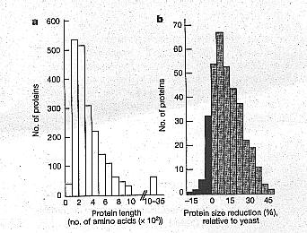 |
| Figure 2 Sizes of E. cuniculi (EO proteins and comparison with S. cerevisiae (Sc) homologues. a. Distribution of the lengths of all the potential E cuniculi protein chains. Only six haye more than 2,000 amino acids (maximum 3,456 amino acids). b. Degrees of reduction in length of E. cuniculi proteins (n = 350) relative to those of S. cerevisiae, expressed as a percentage: 100 (Sc protein length - Ec protein length)/(Sc protein length). The positive classes representative of shorter E. cuniculi proteins are in grey. The dynein heavy chain, the largest protein chain with a clearly predicted function (3,151 amino acids), Is 23% shorter than the yeast homologue (4,092 amino acids). Mean values associated with major functional categories are 7.3% for 'protein synthesis', 11.0% for 'Protein destination', 11.4% for 'metabolisrn/energy', 14.4% for 'intracellular transport 15.3% for 'transcription' and 20.1% for 'cell growth, cell division and DNA synthesis'. |
The present genome sequence extends this evidence: phylogenetic analyses of putative genes for seryl-tRNA synthetase, transcription initiation factor IIB, subunit A of vacuolar ATPase, and a GTP-binding protein place microsporidia as a sister group of fungi with bootstrap supports ranging from 70% to 92% (see Supplementary Information; a systematic phylogenetic analysis of the genome will be presented elsewhere).
Figure 3 An overview of metabolism and transport in E. cuniculi, as deduced from genome sequence analysis. Pathways for nucleotide biosynthesis, energy production and chitin biosynthesis are indicated. Endocytosis and vesicular transport involving a cis-trans polarized Golgi apparatus are also illustrated. Potential transporters associated with the plasma membrane are shown with indications on substrate specificity. Question marks correspond to major uncertainties about the fate of pyruvate and the production of second messengers for signal transduction. The parasite is represented within a parasitophorous vacuole (PV) of the host cytoplasm.

Genes of putative mitochondrial
evolutionary origin in the E. cuniculi genome were systematically sought
by comparison with the 423 recently surveyed yeast mitochondrial proteins
encoded by the nuclear and the mitochondrial genomes (22). Twenty-two genes
with significant similarity to the yeast genes were identified and phylogenetic
analysis showed that six of them are closely related to homologues from alfa-proteobacteria,
the bacterial group from which mitochondria are believed to derive (23). The
yeast homologues of these six proteins are ATM1 (ABC transporter), ISU1/ISU2
(NIFU-like protein), NFS1 (unique homologue of bacterial ISC-S and NIF-S),
SSQ1 (heat-shock protein of relative molecular mass 70,000), YAH1 (ferredoxin)
and PDB1 (beta-subunit of pyruvate dehydrogenase component E1). The first
five proteins are typically involved in the Fe-S cluster assembly machinery~
an essential function of mitochondria (24).
The presence of characteristic domains and key amino acids suggests that the
potential mitochondrial-type proteins are functionaL Moreover, PSORT analysis
predicts amino-terminal presequences for the targeting of five of these proteins
(see Supplementary Information). A common feature is an arginine residue at
-2 relative to the cleavage site, similar to presequences of mitochondrial
and hydrogenosomal proteins (25). The amitochondriate protozoan Entamoeba
histolytica has recently been shown to contain a residual mitochondrion-derived
organefle (26,28) that some authors have called mitosome (28). Considering
the set of potential E. cuniculi proteins usually associated with mitochondria,
we propose that a cryptic organelle is also present in microsporidia (Fig.
4). This mitosome would be significantly different from hydrogenosomes found
in several anaerobic unicellular eukaryotes (type II anaerobes) (29). Hydrogen
production through pyruvate catabolism seems unlikely in microsporidia (because
of a lack of a hydrogenase gene). The development of microsporidia in various
aerobic host cells is suggestive of a rather high 02
tolerance and therefore of an efficient protection against oxidative stress.
No catalase gene is identified, in agreement with the lack of peroxisomes.
Thus, in addition to glutathione and thioredoxin-based systems, E. cuniculi
might use its unique manganese superoxide dismutase as an antioxidant.

Figure 4 Conceptual scheme of a mitochondrion-derived organelle ('mitosome') in E. cuniculi suggested by the detection of several homologues of mitochondrial proteins. Under this hypothesis, pyruvate decarboxylation may occur through a heterotetrameric form (alfa2beta2) of the pyruvate dehydrogenase E1 component (PDH-E1) and transfer of reducing power towards the organelle transits through a glycerol -3-phosphate shuttle involving both cytosolic (GPDH-C) and mitochondrial (GPDH-M) glycerol-3-phosphate dehydrogenases. By analogy with hydrogenosomal pyruvate:ferredoxin oxidoreductase, a system based on ferredoxin (Fdx) and NAD(P)H ferredoxin:oxidoreductase (FOR) is assumed to be used for acetate production. A cytosolic acey coenzyme A (CoA) synthetase (ACS1) may catalyse acetate activation. Considering an aerobic environment and the lack of hydrogenase, a simplified but specific electron transport towards molecular oxygen remains a possibility. The predicted manganese superoxide dismutase (Mn-SOD) would ensure protection against oxygen radicals. A homologue of yeast ERV1 (a small protein required for mitochondrial biogenesis) is indicated. In the lower part of the scheme are depicted some of the major potential factors required for targeting of proteins to the organelle and for biosynthesis of Fe-S clusters and transport of Fe-S proteins towards the cytosol. Iron is predicted to be essential for controlling the expression of mitosomal proteins.
This first report of the genome sequence of a eukaryotic parasite should stimulate
proteomic approaches to identify gene products of interest for diagnosis and
therapy of microsporidioses, as well as to test the mitosome hypothesis. In
addition, we expect the E. cuniculi genome to provide a useful reference
for comparative genomics of microbial eukaryotes, particularly to identify
the relative importance of shared genes among various evolutionarily distant
intracellular parasites, including major human-infecting parasites such as
Plasmodium and Leishmania.
Methods
The reference mouse isolate of Encephalitozoon cuniculi (GB-M 1), cloning
and library construction, nudeotide sequencing, sequence validation and sequence
analysis have been described in detail previously (11)(see Supplementary Information).
Further information on recombinant DNA preparation, sequencing and sequence
analysis can be found elsewhere (30). Annotation was manually performed with
the help of AceDB and Artemis graphic interfaces. Genes were characterized
by Glimmer prediction of coding DNA sequences, combined with BLAST all-homology
results (BLAST X, BLAST N against 'nr', BLAST P against SwissProt, PSI-BLAST).
Transfer RNA genes were detected with the tRNA Scan program. Spliceosomal-type
introns were manually detected. Phylogenetic analyses were done as in Keeling
et al. (8). Gamma-corrected ML distances with eight rate categories and invariant
sites were computed with TREE-PUZZLE version 5.0 and trees were derived with
BioNJ. Bootstrapping was on 500 replicates with alpha parameters and the fraction
of invariant sites estimated once from the original data. Sequences of individual
chromosomes were submitted to EMBL under the accession numbers AL39173 for
chrI and AL590442-AL590451 for chrII-chrXI, respectively.
Received 23 April; accepted 31 August 2001.
1. Wittner, M. & Weiss, L. M. The Microsporidia and Microsporidiosis
(American Society of Microbiology, Washington DC, 1999).
2. Vossbrinck, C. R., Maddox J. V, Friedman, 5., Debrunner-Vossbrinck, B.
A. & Woese, C. R.
Ribosomal RNA sequence suggests microsporidia are extremely ancient eukaryotes.
Nature 326,411414(1987).
3. Germot, A., Philippe, H. & Le Guyader, H. Evidence for loss oftnitochondria
in microsporidia from a mitochondrial-type HSP70 in Nosema locustae. MoL Biochem.
Parasitol. 87, 159-168 (1997).
4. Hirt, R. R, Healy, B., Vossbrink, C. R., Canning, E. U. & Embley T.
M. A mitochondrial Hsp70 orthologue in Vairimorpha necatrir. molecular evidence
that microsporidia once contained mitochondria. Curr. BioL 7, 995-998 (1997).
5. Peyretaillade, E. et al. Microsporidia, amitochondrial protists, possess
a 70-kDa hut-shock protein gene of mitochondrial evolutionary origin. MoL
BioL Evol. 15, 683-689 (1998).
6. Hirt, R. P. et al. Microsporidia are related to fungi: evidence from the
largest subunit of RNA polymerase II and other proteins. Proc. Natl Acad Sci.
USA 96, 580-585 (1999).
7. Baldauf, S. L., Roger, A. I., Wenk-Siefert, 1. & Doolittle, W. F. A
kingdom-level phylogenyofeukaryotes based on combined protein data. Science
290, 972-977 (2000).
8. Keeling, P. J., Luker, M. A. & Palmer, J. D. Evidence from 0-mbulin
phylogeny that microsporidia evolved from within the Fungi. Mol. Biol Evol
17, 23-31 (2000).
9. Van de Peer, Y., Ben Ali, A. & Meyer, A. Microsporidia: accumulating
evidence that a group of
amitochondriate and suspectedly primitive eukarymes are just curious fungi.
Gene 246, 1-8 (2000).
10. Biderre, C., Pagh, M., Mitinier, G., Canning, F- U. & Vivarb, C. P.
Evidence for the smallest nuclear genome (2.9 Mb) in the microsporidian Encephalitozoon
cuniculi. MaL Biochem. Parasitol. 74, 229231 (1995).
11. Peyret, P. et al. Sequence and analysis of chromosome I of the amitochondriate
intraceflular parasite Encephalitozoon cuniculi (Microspora). Genome Res.
11, 198-207 (2001).
12. Brugère, J.F., Cornillot, L, Méténier, G., Bensimon,
A. & Vivaris, C. P. Encephalitozoon cuniculi (Microspora) genome: physical
map and evidence for telomere-associated rDNA units on all chromosomes. Nucleic
Acids Res. 28, 2026-2033 (2000),
13. Douglas, 5. et al. The highly reduced genome of an enslaved algal nucleus.
Nature 410, 1091-1096 (2001).
14. Zitang, L Protein-length distributions for the three domains of life.
Trends Genet. 16,107-109 (2000). Shiralcura, T. et al. Characterization ofthe
ribosomal proteins ofthe amitochondriate protist Giardia lamblia. MoL Biochem.
Parasitol. 112,153-156 (2001).
16. Spingola, M., Grate, L., Haussler, D. & Ares, M. Genome-wide bioinformatic
and molecular analysis of introns in Saccharomyces cerevisiae. RNA 5, 221-234
(1999).
17. El Alaoui, H., Bata, J., Bauchart, D., Dori, J.-C. & Vivarb, C. P.
Lipids of three microsporidian species and multivariate analysis of the host-parasite
relationship. 1. ParasitoL 87, 554-559 (200 1).
18. Weidner, L, Findley A. M., Dolgikh, V. & Skoloya, J. in The Microsporidia
and Microsporidiosis
(eds Wittner, M. & Weiss~ L. M.) 172-195 (American Society ofMicrobiology,
Washington DC, 1999).
19. Wolf, Y. L Aravind. L & Koonin, E. V. Rickettsiae and Chlamydiae..
evidence of horizontal transfer and exchange. Trends Genet 15, 173-175 (1999).
20. Böhne, W., Ferguson, D. j., Kohler, K. & Gross, U. Developmental
expression ofa tandemly reputed, glycine- and serine-rich spore wall protein
in the microsporidian pathogen Encephalitozaon cuniculi. Infect. Immun. 68,
2268-2275 (2000).
21. Delbac, E, Peuvel, L, Wtinier, G., Peyretaillade, E. & Vivarès,
C. P. Microsporidian invasion apparatus: identification of a novel polar tube
protein and evidence for clustering of ptp1 and ptp2 genes in three Encephalitozoon
species. Infect. Immun. 69,1016-1024 (2001).
22. Karlherg, 0., Canback, B., Kurland, C. G. & Andersson, S. G. The dual
origin of the yeast mitochondkial proteome. Yeast 17.170-187 (2000).
23. Andersson, 5. G. et aL The genome sequence ofRickettsia prowazekii and
the origin ofrititochondria. Nature 396,133-140 (1998).
24. Lill, R. & Kispal, G. Maturation ofcellular Fe-S proteins: an essential
function of mitochondria. Trends Biochern. Sci. 25, 352-356 (2000).
25. Dyall, S. D. & Johnson, P. 1. Origins of hydrogenosomes and mitochondria:
evolution and organelle biogenesis. Curt. Opin. MicrobioL 3, 404-411 (2000).
26. Rodriguez, M. A. et aL The pyruvate ferredoxin oxidoreductase is located
in the plasma membrane and in a cytoplasmic structure in Entamoeba. Microb.
Pathog. 25, 1-10 (1998).
27. Mai, Z. et at Hsp60 is targeted to a cryptic mitochondrion-derived organelle
('crypton') in the micr aerophilic protozoan parasite Entarnoeba histolytica.
MoL CelL BioL 19,2198-2205 (1999).
28. Tovar, J., Fischer, A. & Clark, C. G. The mitosome, a novel organelle
related to mitochondria in the amitochondrial parasite Entamoeba histolytica.
Mol. Microbial, 32,1013-1021 (1999).
29. Martin, W. & Miffier, M. The hydrogen hypothesis for the first eukaryme.
Nature 392, 37-41 (1998).
30. Artiguenave, F. et al. Genomic exploration of the herniascomycetous yeasts:
2. Data generation and processing. FEBS Lett, 487, 13-16 (2000).
Supplementary Information
accompanies the paper on Nature's website
(http://www.nature.com).
Acknowledgements
We are grateful to everyone who provided computer resources and system management:
D. Ferney and P. Bernard (LMA, UMR CNRS 6620), P. L. Reichstadt and B. Michel
(LPC, IN2P3, UMR CNRS 6533). We thank B. Chebance and R. Guerry for technical
assistance.
L. Pereira, B. Duperrier, R. Météry and E. Delorme are acknowledged
for their contribution to the development of the annotation environment in
the Université Blaise Pascal. We also express gratitude to C. White
for critical reading of the manuscript. FD, H.E.A and E.P were supported by
a grant from the Ministère de la Recherche.
Correspondence and requests for materials should be addressed to C.P.V.
(e-mail: christian.vivares@lbp.univ-bpclermont.fr).
da NATURE ‡ VOL 414 ‡ 22 NOVEMBER 2001 ‡ www.nature.com